

17 Feb 20
A patient with severe heart failure had a resting SpO2 values at the lower limit of normal , but had brief episodes of desaturation ( SpO2 falls ) as if hypo ventilated.
He also reported wheezing for minimal effort and episodes of nocturnal awakenings with a sense of suffocation.
He performed a 3D oximetry test during his sleep with a particular oximeter equiped with an accelerometer that allows the phases of movement (ie awake) to be distinguished by presumed sleep periods and to identify the positions taken during the sleep just as a polysomnography does.
The result was surprising: at first glance the plot suggested the presence of typical rapid desaturation sleep apnea syndrome (OSAS ).
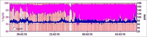
Fig. 1 - Sleep oximetry : tracing the entire night.
However, performing the analysis by position something particularly sensational emerged. In the early hours of recording the patient was awake (as he confirmed), in motion (the accelerometer has registered more than 800 paths distance traveled).
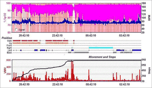
Fig. 2 - Sleep oximetry: complete analysis of the position and movement (paths).
Rapid and serious desaturation occurred not only during the sleep but also while he was awake and walking.
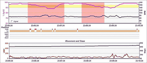
Fig. 3 - Most particular whilst the subject is awake and standing on his feet and walking.
In addition to the desaturation a cyclical movement called periodicity was shown with an asymmetrical look different from that of OSAS.
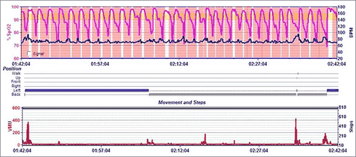
Fig. 4 - Sleep oximetry: partial tracing that shows the periodicity of desaturation.
In this case the descent of the desaturation occurs in several stages: first slow descent along the plateau due to hypoventilation and second rapid descent due to an almost cessation of flow ( apnea). Finally, the subsequent slow ascent due to the gradual resumption of normal ventilation.
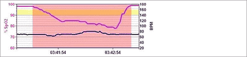
Fig. 5 - Sleep oximetry: an example of an episode of desaturation due to Cheyne Stokes Respiration.
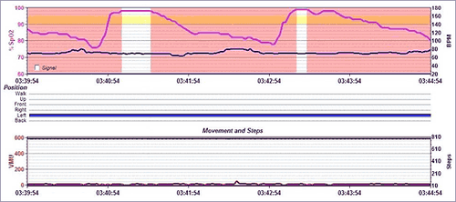
Fig. 6 - Sleep oximetry: a sequence of desaturation of the subject under consideration due to Cheyne Stokes Respiration.
In the case of sleep apnea the desaturation has instead a single phase of prolonged and continuous descent to the point of minimum SpO2 while the subsequent ascent is short and steep because of the resumption of breathing.
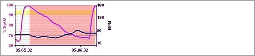
Fig. 7 - Sleep oximetry: an example of an episode of desaturation due to sleep apnea (OSA).
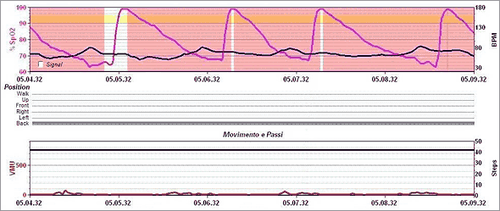
Fig. 8 - Sleep oximetry: a sequence of desaturation caused by sleep apnea (OSA).
To ensure that oxygen gets to the cells, it is necessary that it goes through breathing from the atmosphere to the lungs alveoli through the upper airways and bronchi with a cyclic movement of the chest (ventilation ) .
This movement is controlled by the respiration centre of the brainstem and and mobilizes approximately 0.5 liters of air per breath at rest ( tidal volume ) for about 12 breaths per minute ( respiratory rate ) .
In a healthy subject at rest the breathing rhythm is very regular . Even in many obstructive respiratory diseases (asthma and COPD) or restrictive ( fibrosis ) in which you change the current volume the breathing rhythm is maintained regularly.
However, with periodic breathing there is an irregular rhythm even if periodic. The most popular model is the so-called Cheyne Stokes breathing , present in heart failure , probably due to the increase of the transit time of blood from the lungs to the Center of the Breath .
This model is characterized by the continuous repetition of periods of progressive decrease in tidal volume followed by a short period of apnea and an increase in tidal volume , as described above. This sequence is repeated with remarkable regularity.
Bibliography:
Olson EJ, Moore WR, Morgenthaler TI et al. Obstructive Sleep Apnea-Hypopnea Syndrome. Mayo Clin Proc. 2003; 78: 1545-52.
Mbata GC and Chukwuka JC. Obstructive Sleep Apnea Hypopnea Syndrome. Ann Med Health Sci Res. 2012; 2: 74–77.
Quaranta AJ, D'Alonzo GE, Krachman SL. Cheyne-Stokes respiration during sleep in congestive heart failure. Chest. 1997 ; 111(2): 467-73.
AlDabal L, BaHammam AS. Cheyne-stokes respiration in patients with heart failure. Lung. 2010; 188(1): 5-14.
Yumino D, Bradley TD. Central sleep apnea and Cheyne-Stokes respiration. Proc Am Thorac Soc. 2008 Feb 15;5(2):226-36.
Pino G, Milone PA, Ciappi G. Ipoventilazione alveolare e iposensibilità dei centri respiratori in un caso di sindrome diencefalica. Rassegna di Patologia dell’Apparato Respiratorio 1963-, 13 (4): 3-18.
Farré R, Montserrat JM, Navajas D. Noninvasive monitoring of respiratory mechanics during sleep. Eur Respir J. 2004; 24(6): 1052-60.
Gastaut H, Tassinari CA, Duron B.Polygraphic study of the episodic diurnal and nocturnal (hypnic and respiratory) manifestations of the Pickwick syndrome. Brain Res. 1966 ; 1(2): 167-86.
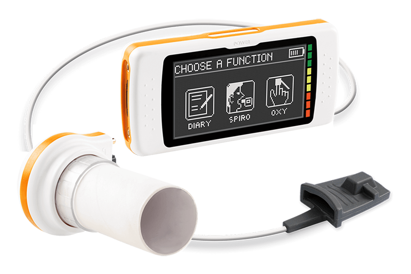
Touchscreen Spirometer with Optional 3D Oximeter


