
16 Apr 24
Spirometry is a lung function test that can be used to detect, follow, and manage lung diseases, as well as evaluate the efficacy of treatment plans. Thanks to technology advancements, spirometry results are now much more reliable than in the past, so long as the healthcare provider knows how to interpret and put them in comparison with the appropriate reference values.
In this article, we will guide you through the basic steps toward the proper interpretation of spirometry results.
During a spirometry test, patients will be required to take a deep breath and then exhale into a device named a spirometer in order to measure and record a series of parameters related to lung function and performance, like the amount of air they can breathe in and out, or how fast and steadily they can exhale.
The three main indices needed for an accurate evaluation of a patient’s pulmonary health are:
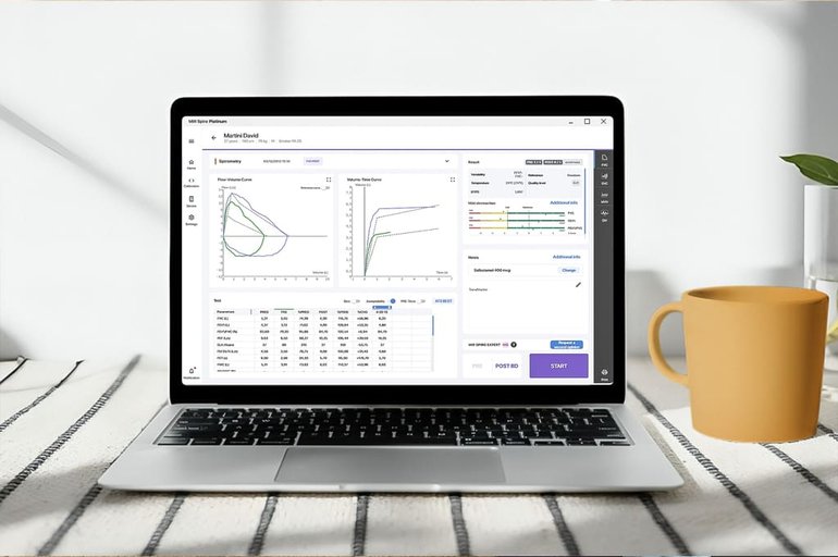
Most spirometers will also provide a visual record of the expiration in the form of a graph known as a spirogram. The breath flow will usually be shown on the X axis and expressed in lt/s, while the breath volume will be shown on the Y axis and expressed in lt.
In order for spirometry results to be accurate and acceptable, it is always advisable to follow a series of best practices related to both spirometer maintenance and patient education.
Elements like glottis closure, cough, early termination or submaximal effort from the patient, as well as leaks or obstructed mouthpieces can generate artifacts in your visual results, invalidating the test.
Your spirometer must be calibrated based on the patient’s history and demographic (age, gender, height, etc.). This will help you select the appropriate reference equation, which is derived from healthy, nonsmoking subjects sharing similar demographics, and will provide the reference values for your test.
A minimum of three acceptable tests should be obtained for appropriate evaluation of a patient’s lung condition.
Recorded values are usually shown in columns and compared to the normal range, also called expected values, in the form of a percentage. If the values measured fall over 80% of the predicted score, the result is considered normal, which generally means a patient’s lungs are functioning well, while lower percentages are indicators of respiratory issues.
If the values fall above the normal range, there are usually no clinical consequences. However, if the recorded parameters are significantly higher than normal, you may want to check your spirometer calibration or make sure you selected the correct reference values.
As anticipated before, normal spirometry results will show FVC, FEV1 and FEV1/FVC values that fall within the normal range. If the values fall below the expected 80%, thus indicating the presence of respiratory issues, it will be necessary to identify whether we are in the presence of obstructive or restrictive patterns.
Obstructive patterns are characterized by low FEV1/FVC ratio, usually below 70%.
This happens because there are obstructions in the airways that prevent the patient from breathing out most of the inhaled air during the first second of forced expiration. Obstructive patterns are therefore usually accompanied by low FEV1 – though there may be exceptions – and are often indicators of conditions like asthma or COPD.
Restrictive patterns are characterized by low FVC and FEV1 values, with a normal FEV1/FVC ratio, indicating that your lungs cannot fully expand during inspiration. This may be due to restrictive respiratory conditions, such as pulmonary fibrosis or scoliosis. In this case, it is advisable to confirm restriction by measuring the lung volumes and total lung capacity.
If you need to know more about normal values in spirometry, we explored the topic in this article.


