
19 Oct 26
Two patients with COPD may have the same FEV₁ yet markedly different pathophysiological and structural characteristics.
Quantifying airflow limitation is fundamental, but it does not, on its own, capture the full heterogeneity of the disease. The shape of the flow–volume curve contains additional information, and the Emphysema Severity Index (ESI) was developed to express this information quantitatively.
When interpreting spirometry, pulmonologists naturally examine FEV₁, FVC and FEV₁/FVC. They also examine the shape of the expiratory portion of the flow–volume curve.
Clinical experience has taught us to associate increasing concavity of the curve—particularly when its trajectory departs from the normal, approximately mono-exponential decline in flow—with an emphysematous component and small airway involvement.
The difficulty is that this assessment has traditionally been visual, qualitative and inevitably subjective.
ESI is based on a very simple concept: converting this visual observation into a quantitative measure.
ESI describes the shape of the expiratory portion of the flow–volume curve using a continuous score ranging from 0 to 10.
The calculation uses five measurements already available from conventional spirometry:
In other words, ESI assigns a numerical value to a feature of the curve that experienced clinicians are already accustomed to assessing visually.
A distinctive feature of the index is that it is calculated using absolute spirometric values. It therefore requires no predicted values, adjustments for age, sex or height, or population reference equations.
This is probably the most clinically interesting aspect.
Two patients may have the same degree of FEV₁ reduction but different underlying pathophysiological mechanisms.
For a given degree of airflow limitation, a high ESI makes a substantial emphysematous component highly likely, associated with loss of elastic recoil and its effects on the small airways.
A relatively low ESI, by contrast, points towards a greater contribution from disease in the more proximal airways, as may occur in the chronic bronchitic phenotype.
FEV₁ and ESI therefore provide different, complementary information: FEV₁ primarily quantifies the severity of airflow obstruction, while ESI helps describe the pathophysiological mechanism underlying the particular shape of the expiratory curve.
COPD is a heterogeneous disease. Patients with an apparently similar degree of airflow obstruction may have different phenotypes.
The ability to better characterise a predominantly emphysematous component versus a predominantly bronchial component may, alongside symptoms, clinical history and other available data, contribute to a more individualised patient assessment and potentially to more appropriate tailoring of treatment choices.
ESI should therefore be interpreted in conjunction with the other elements of the clinical and functional assessment, rather than in isolation.
The relationship between ESI and structural lung abnormalities has been investigated using CT.
Initial studies demonstrated associations between ESI and the presence and severity of emphysema assessed by quantitative CT (Occhipinti M, et al. 2019). Subsequent validation in 5,930 participants in the COPDGene study, who underwent spirometry and CT on the same day, confirmed that the shape of the expiratory curve can yield physiological information related to the structural component of the disease (Occhipinti M, et al. 2020).
This does not mean that ESI can replace CT when anatomical characterisation is required.
CT and ESI assess the disease from different perspectives: CT directly depicts structural abnormalities, whereas ESI describes their expression in expiratory physiology.
The advantage is that this information is obtained from the same spirometric manoeuvre, without radiation exposure, additional investigations or any increase in testing time.
The available evidence suggests that the potential value of ESI may extend beyond patients already diagnosed with COPD.
In a Swedish general population, increasing ESI values were associated with all-cause and respiratory mortality (Luoto J, et al. 2022).
More recently, an Italian epidemiological study showed that ESI can also provide clinically meaningful information in individuals without airflow limitation according to conventional spirometric criteria (Maio S, et al. 2026). These findings suggest potential value in the assessment of smokers and individuals at risk.
Of course, an elevated ESI in an individual without airflow limitation does not, on its own, constitute a diagnosis of COPD or emphysema: it provides additional physiological information that must be interpreted in the clinical context.
A further development is being investigated in a manuscript currently in preparation, in which ESI was used in place of CT criteria within a multidimensional framework for COPD diagnosis (Castaldi PJ, et al. 2026).
The main finding indicates that, when CT is unavailable, ESI can capture some of the information provided by structural CT criteria, particularly in patients with airflow limitation, using only information already contained in conventional spirometry.
This finding should be considered preliminary until the study is published, but it is consistent with the pathophysiological concept underlying the index: using the shape of the expiratory curve as a physiological descriptor of structural lung abnormalities.
For pulmonologists, ESI quantifies information traditionally sought through visual assessment of the flow–volume curve and complements conventional parameters.
For general practitioners, automatic calculation provides additional information to help characterize the patient without requiring a new manoeuvre or a new test.
In both cases, ESI preserves the role of traditional spirometric parameters and does not replace other diagnostic tools when indicated. Rather, it allows fuller use of the information already contained in spirometry.
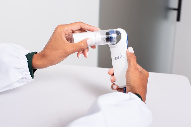
In the Minispir Plus ESI and Spirolab Plus ESI devices, ESI calculation is integrated with standard spirometric parameters.
The distinctive feature is therefore the automatic extraction of additional information from the same manoeuvre, without adding another diagnostic procedure.
No new manoeuvre and no new test: a more complete interpretation of the information already contained in the flow–volume curve.
Discover MIR spirometers with integrated ESI
Ferrara F, De Berardinis F, Scognamiglio M, Zovi A. An Italian regional analysis on chronic obstructive pulmonary disease (COPD): Correct diagnosis and lifestyle play a fundamental role in supporting drug therapy and containing healthcare costs. Naunyn-Schmiedeberg's Archives of Pharmacology 399:20091–20098. https://doi.org/10.1007/s00210-026-05597-y
Maio S, Viegi G, Carrozzi L, Baldacci S, Sarno G, Angino A, Stanisci I, Tagliaferro S, Silvi P, Cenerini G, Paladina M, Occhipinti M, Paoletti M, Pistolesi M, Pistelli F. Emphysema Severity Index: A new spirometric parameter in epidemiology. ERJ Open Research 2026;12:01587-2025. https://doi.org/10.1183/23120541.01587-2025
Luoto J, Pihlsgård M, Pistolesi M, Paoletti M, Occhipinti M, Wollmer P, Elmsthål S. Emphysema severity index (ESI) associated with respiratory death in a large Swedish general population. Respiratory Medicine 2022;200:106899.https://doi.org/10.1016/j.rmed.2022.106899
Occhipinti M, Paoletti M, Bartholmai BJ, Rajagopalan S, Karwoski RA, Nardi C, Inchingolo R, Larici A, Camiciottoli G, Lavorini F, Colagrande S, Brusasco V, Pistolesi M. Spirometric assessment of emphysema presence and severity as measured by quantitative CT and CT-based radiomics in COPD. Respiratory Research 2019;20:101. https://doi.org/10.1186/s12931-019-1049-3.
Occhipinti M, Paoletti M, Crapo JD, Make BJ, Lynch DA, Brusasco V, Lavorini F, Silverman EK, Regan EA, Pistolesi M. Validation of a method to assess emphysema severity by spirometry in the COPDGene study. Respiratory Research 2020;21(1):103. https://doi.org/10.1186/s12931-020-01366-4.
Castaldi PJ, Paoletti M, Occhipinti M Sorano A, Lavorini F, Maiorino E, Hersh CP, Silverman EK, Crapo JD, Bhatt SP, Pistolesi M. The Emphysema Severity Index: Representing CT Abnormalities in a Multidimensional COPD Framework. Manuscript in preparation.


